What Are Oral Tumors?
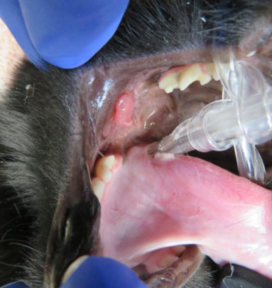
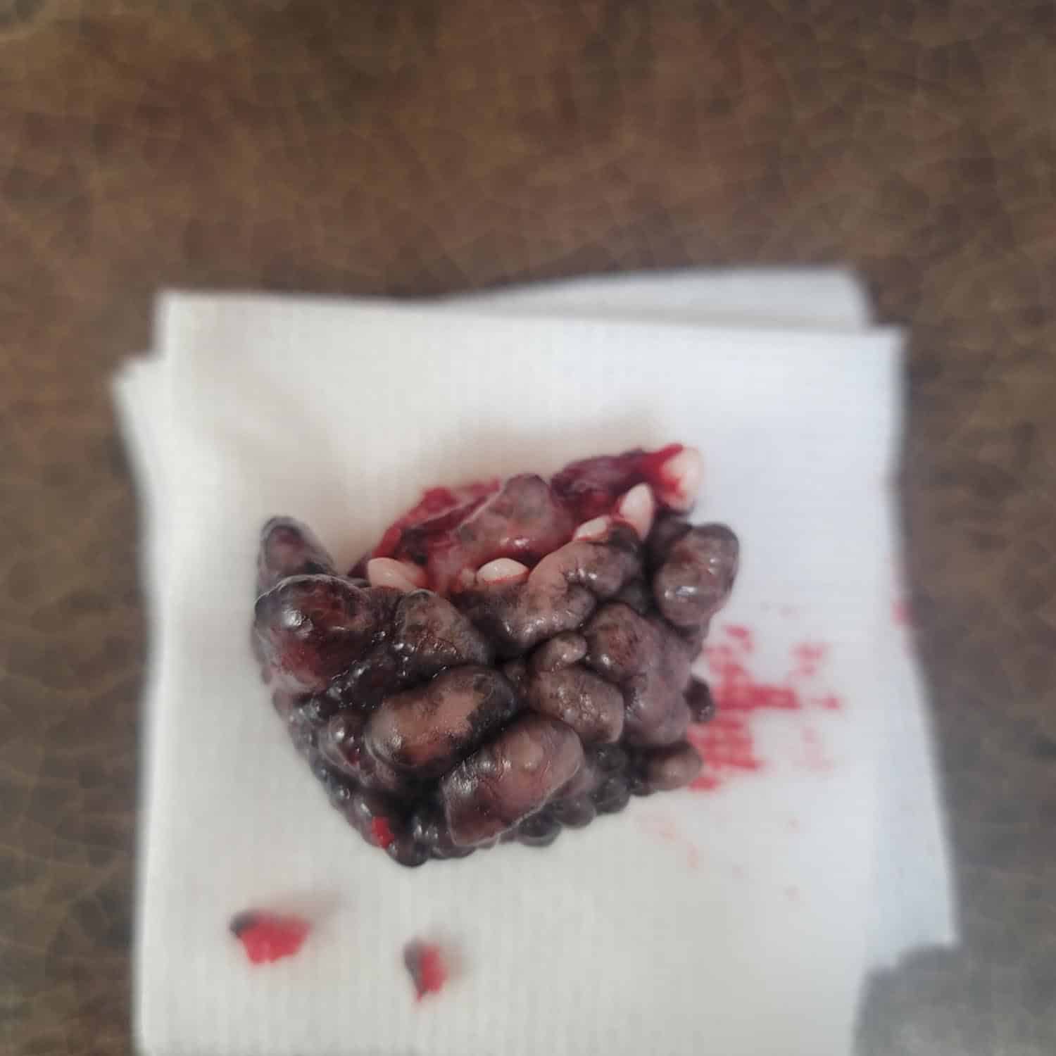
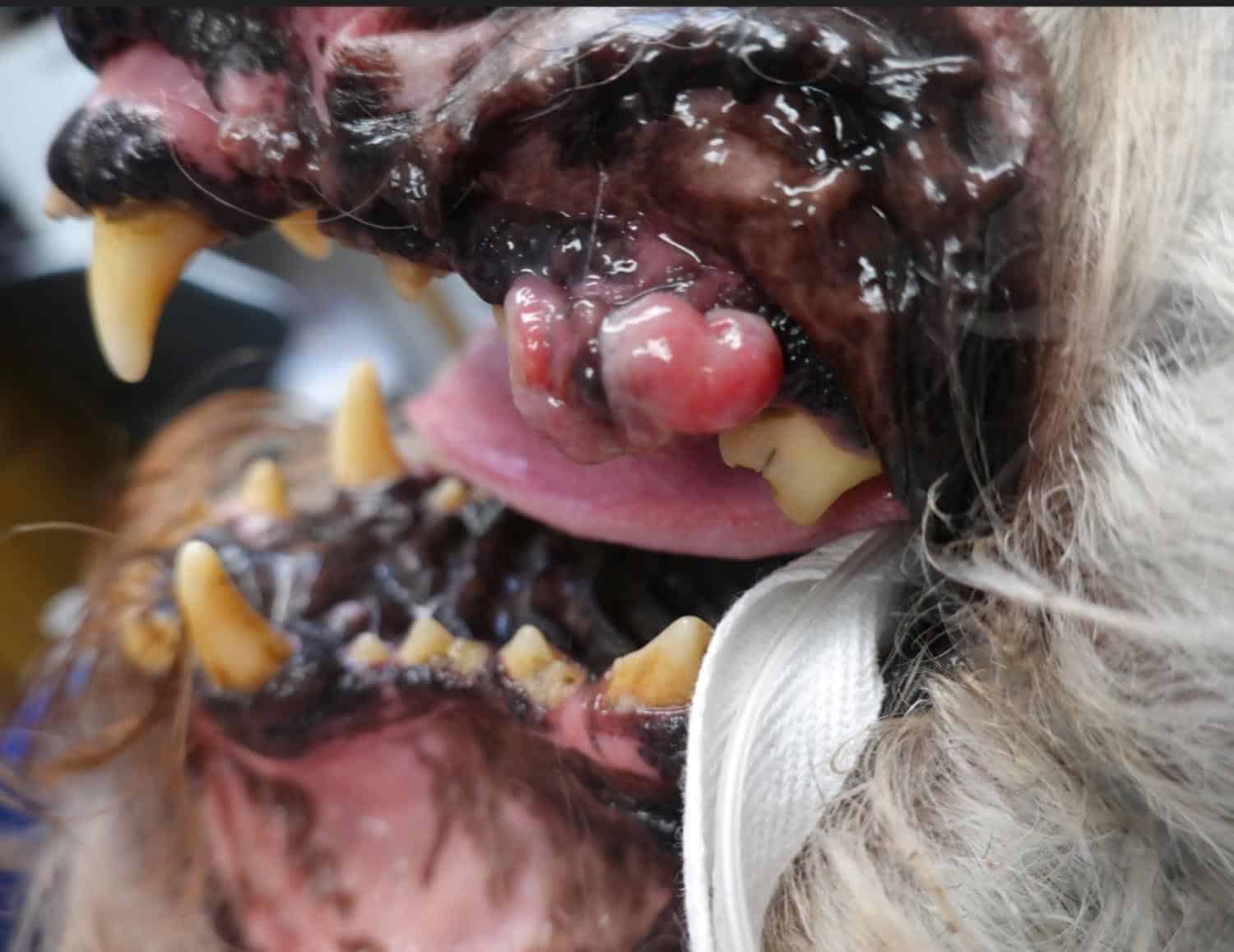
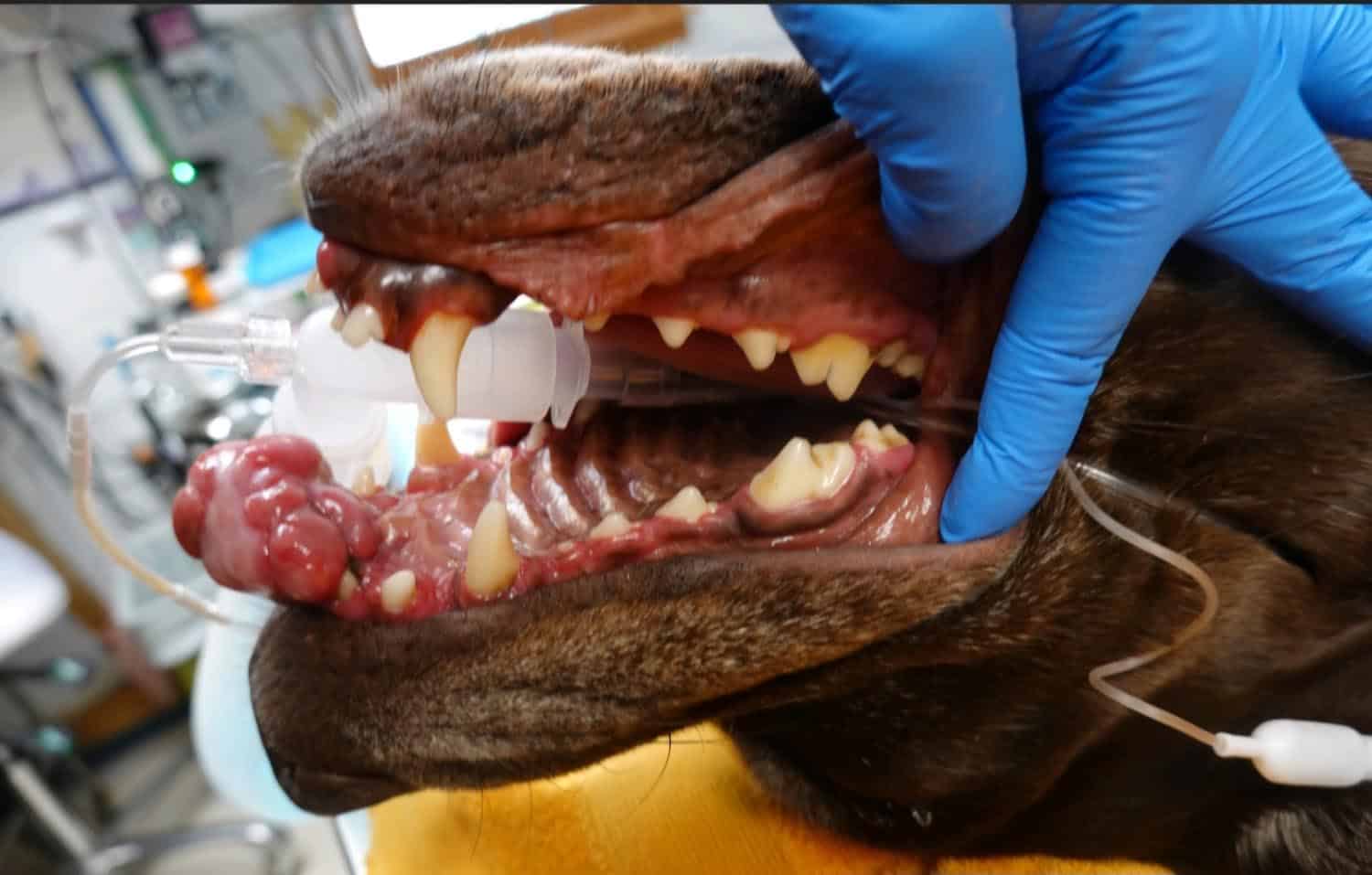
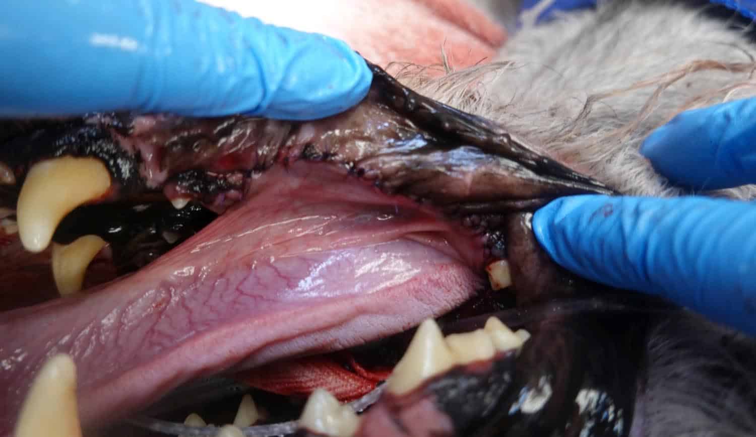
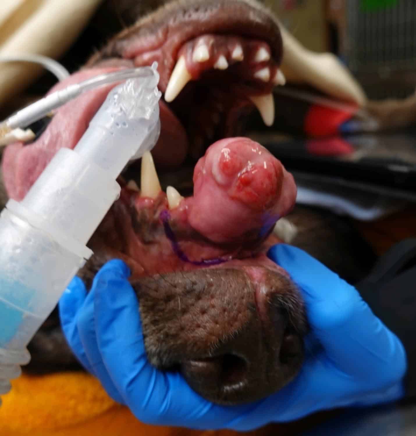
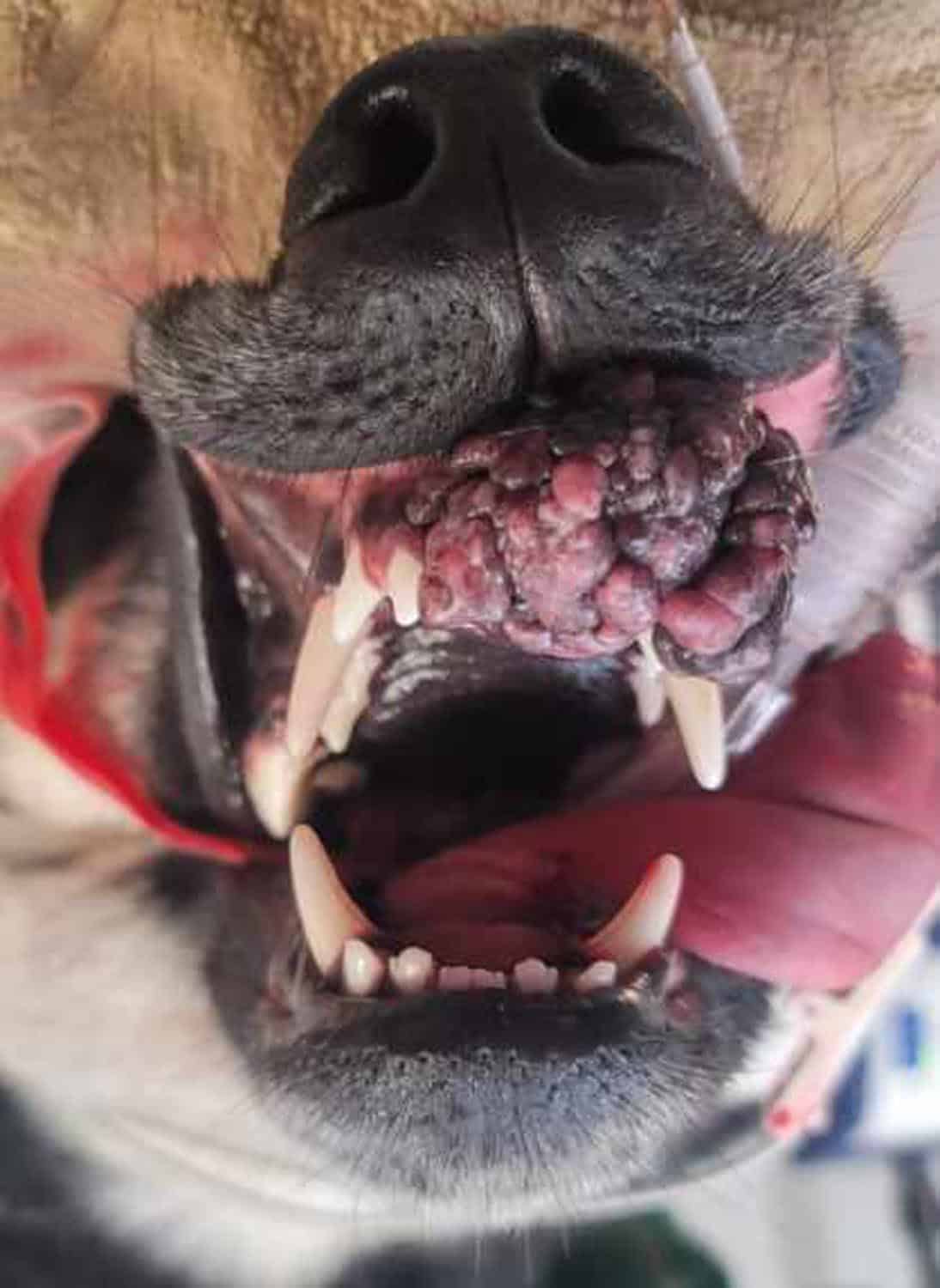
Tumors can occur anywhere in the body and the mouth is no exception. Oral tumors are common in dogs and cats, however, they may not be noted at early stages until the patient is exhibiting symptoms or the owners routinely examine the mouth. Not all tissue enlargements are tumors. An example includes gingival hyperplasia, which is an overgrowth of normal gum tissue, which can lead to “pseudopockets” that can harbor bacteria and cause infection to the teeth. These can be common in brachycephalic breeds like boxers, French bulldogs, pugs, etc.
It is important to diagnose tumors as early as possible. Frequent inspection of the mouth is critical to monitor and detect changes or abnormalities. This can be done by daily tooth brushing to reduce plaque and calculus and aid in maintaining healthy gingiva.
Tumors can be either benign (non-cancerous) or malignant (cancerous). Benign oral tumors do not spread to other parts of the body; however, can be severely locally aggressive. Malignant oral tumors invade the surrounding tissue along with the potential to spread to other parts of the body, particularly local lymph nodes, lungs, or even distant organs. Not all oral tumors present as large growths, some start as non-healing wounds or ulcers.
Assessment and Treatment of Oral Tumors
Intraoral dental radiographs are essential to assess tumor characteristics, in particular the extent the tumor has or has not invaded the bone or surrounding tissue. A careful evaluation to determine the best approach for the patient begins with radiographs. Radiographs may also assist the clinician to determine if the tumor is benign or malignant, although histopathology will provide a clearer picture of the type of tumor and its clinically aggressive or non-aggressive nature. The best way to determine what type of tumor may be affecting your pet’s mouth is by obtaining a biopsy (taking a small portion of tissue that is examined under a microscope) that is reviewed by a boarded oral pathologist. Additional advanced imaging or diagnostics may be recommended to assist in staging. Staging is the ability to determine the extent of cancer, as well as if it has spread to nearby lymph nodes or other parts of the body. Advanced diagnostics, which may be recommended, are computed tomography (CT) to visualize the extent of the tumor as well as can assist with 3D rendering; lymph node aspirates can assist to determine if the tumor has spread to local draining lymph nodes, chest radiographs (x-ray of the lungs), and abdominal ultrasounds. Not all of these diagnostics are always recommended, but it is important to know that these tests may be vital in providing a clearer clinical picture of your pet’s condition and assist in determining the best course of action.
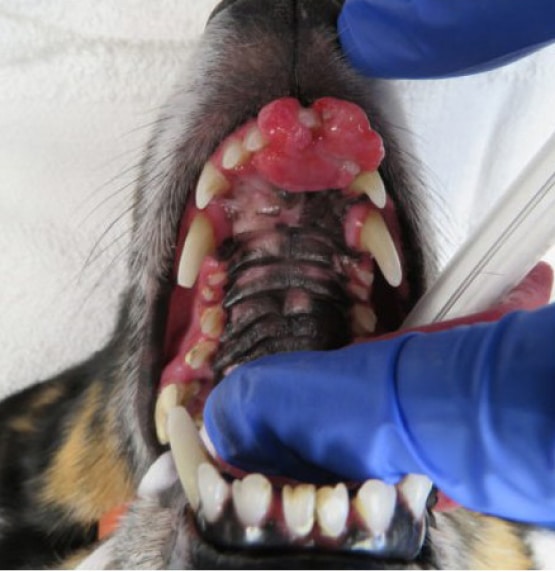
Before
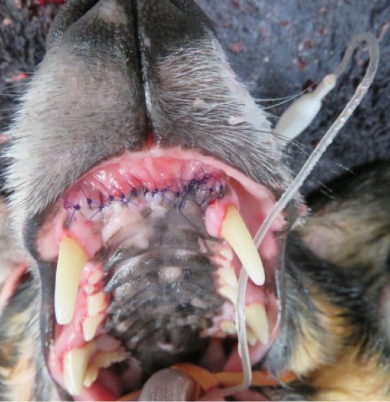
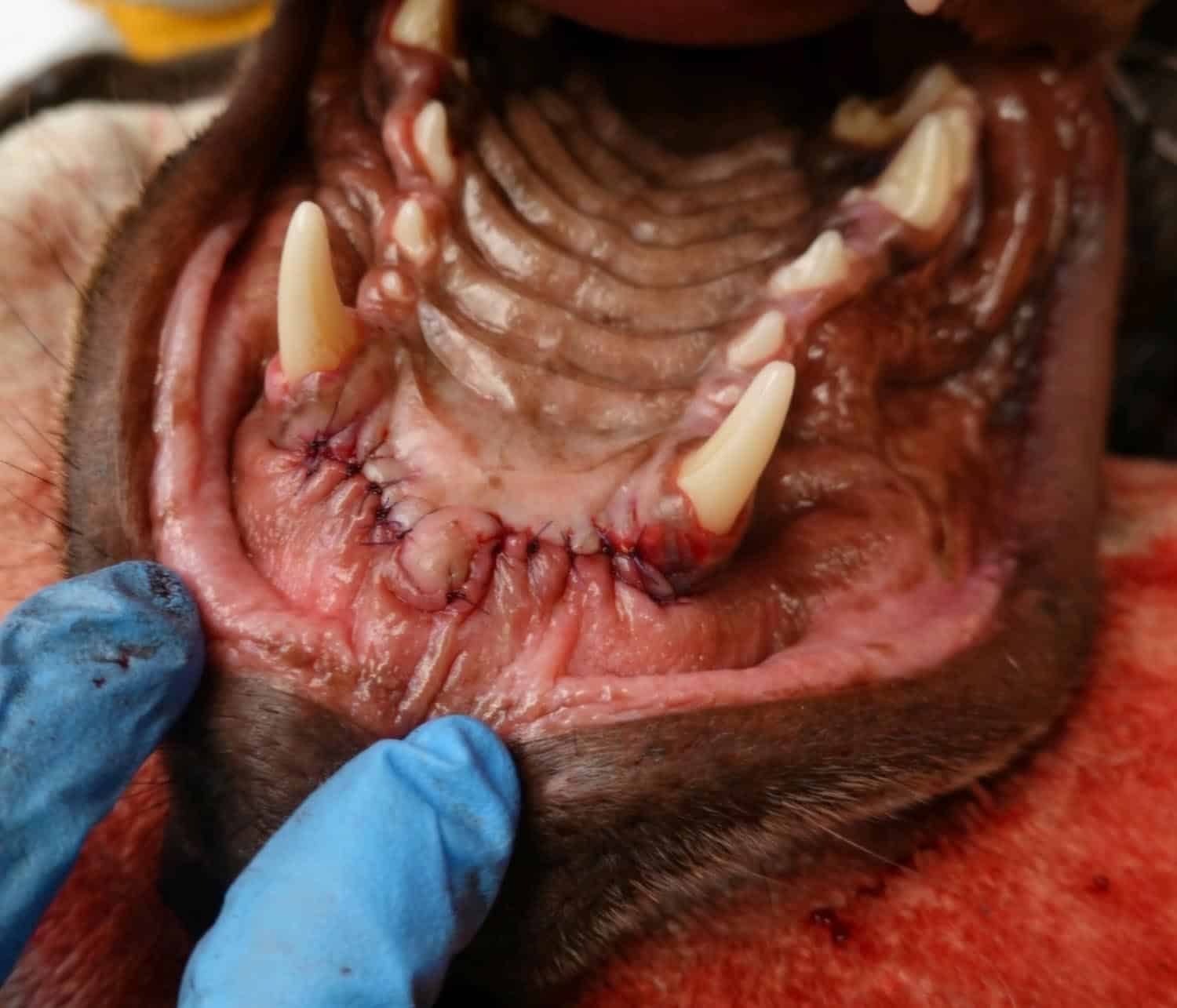
After
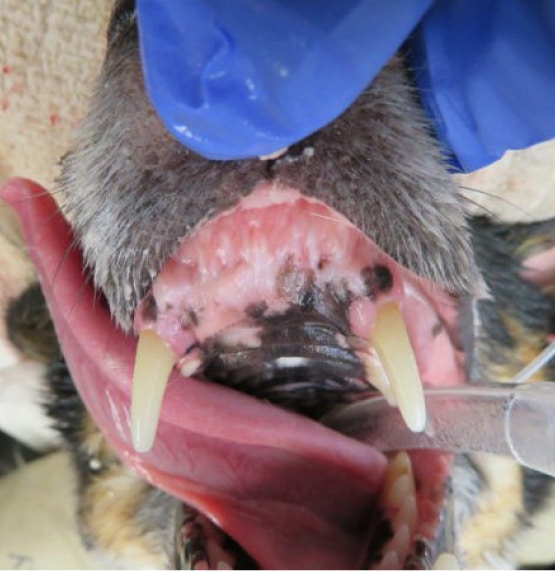
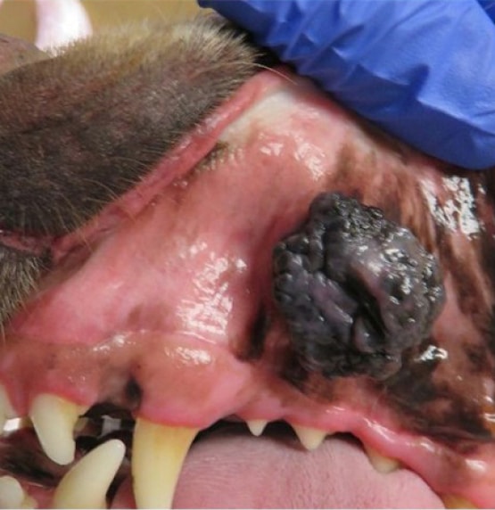
Before
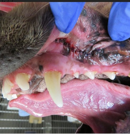
After
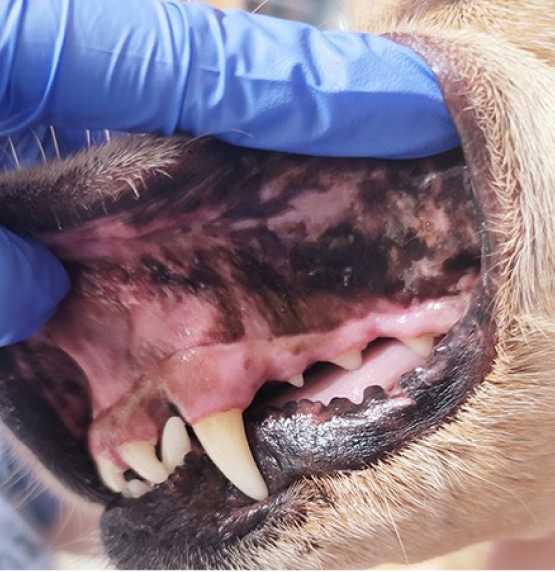
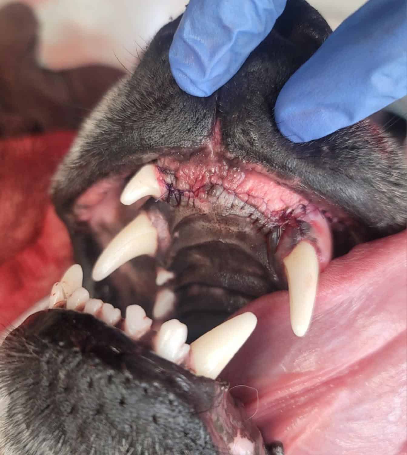
Headline Here
This is just placeholder text. Don’t be alarmed, this is just here to fill up space since your finalized copy isn’t ready yet. Once we have your content finalized, we’ll replace this placeholder text with your real content.
Sometimes it’s nice to put in text just to get an idea of how text will fill in a space on your website.
Traditionally our industry has used Lorem Ipsum, which is placeholder text written in Latin. Unfortunately, not everyone is familiar with Lorem Ipsum and that can lead to confusion. I can’t tell you how many times clients have asked me why their website is in another language!

Call (218) 461-4825 or book online to schedule your pet’s advanced dental appointment.
Frequently Asked Questions About Pet Oral Tumors and Cancer
What are oral tumors in pets, and how are they detected?
Oral tumors are abnormal tissue growths that can occur in the mouths of dogs and cats. Some tumors may not be visible until the pet shows symptoms, which is why regular oral exams and brushing are important for early detection. Not all growths are tumors, and benign conditions like gingival hyperplasia can also affect the gums.
What is staging, and why is it important in the treatment of oral tumors in pets?
Staging is the process of determining the extent of an oral tumor and whether it has spread to nearby lymph nodes or other parts of the body, such as the lungs or abdominal organs. This information is critical for creating an effective treatment plan, as it helps veterinarians assess the severity of the tumor and decide on the best course of action for your pet’s health.
Where do we send all oral pathology samples?
Specialty Oral Pathology for Animals, Dr. Cindy Bell is a Boarded Certified Veterinary Pathologist who is highly specialized in Oral pathology. She has numerous publications in journals and also textbooks. See her website for more information https://www.sopforanimals.com/
After we receive a detailed diagnosis from Dr. Bell what is next for your fur baby?
It is possible surgery alone maybe curative, however, if our specialist has concerns about possible spread, your pet maybe referred to an oncologist. Please know we have your pet’s and your families best interests when we make these recommendations if necessary.
Will my pet need additional surgery?
It maybe recommended to remove local lymph nodes such as the submandibular lymph nodes. If this is the case our Specialist at MNVDS is highly trained at removing these and will walk you through this process if necessary.

